The 人体核磁共振成像核心 Facility is a core dedicated to providing cutting-edge magnetic resonance imaging services for human participants.
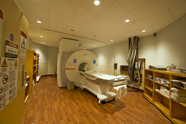
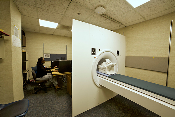
西门子Vida 3T核磁共振扫描仪
PST模拟MRI模拟器
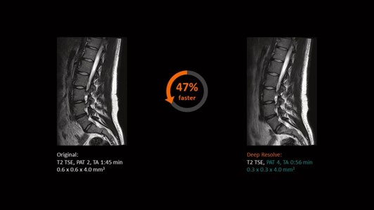
3T西门子Magnetom Vida与生物矩阵技术和涡轮套件加速包
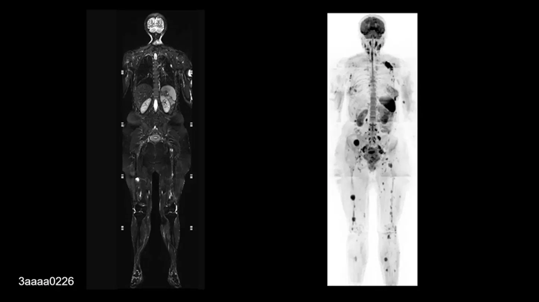
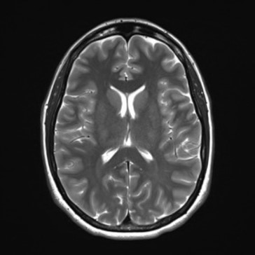
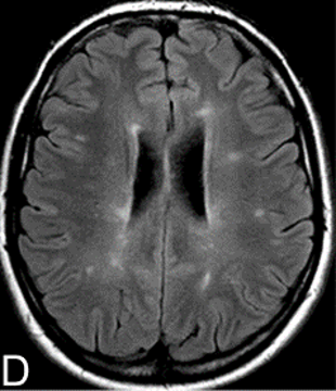
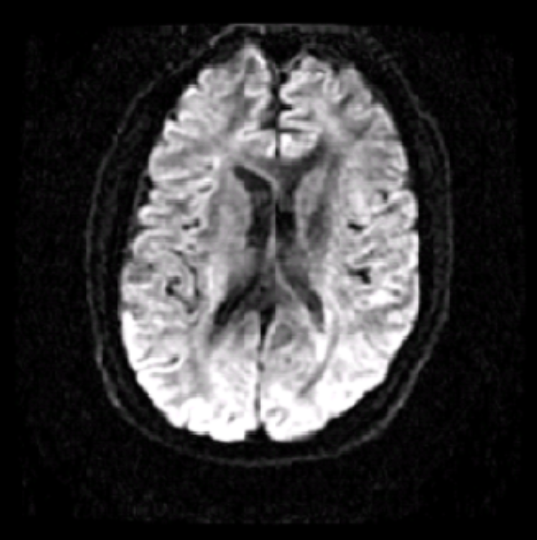
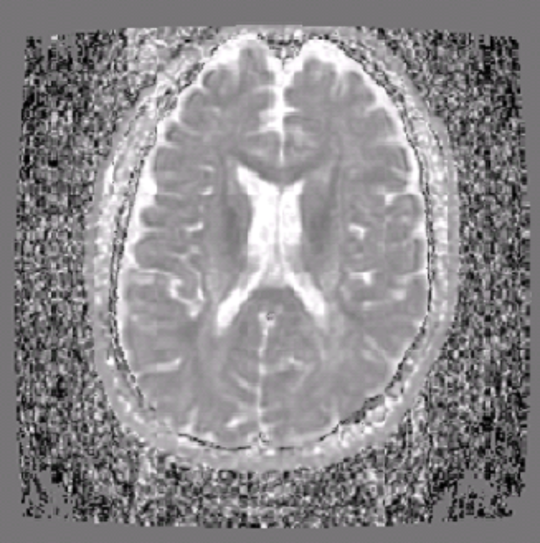
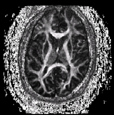
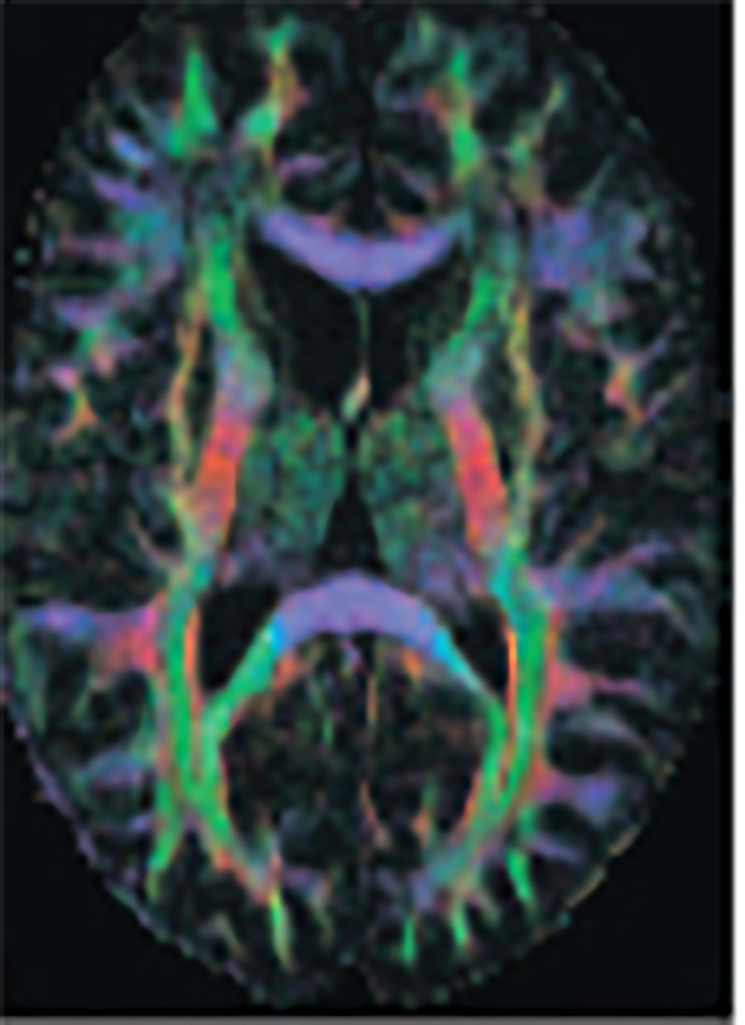
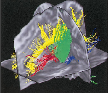
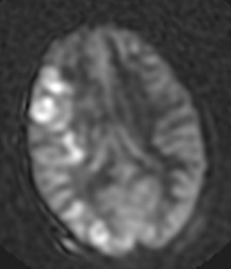
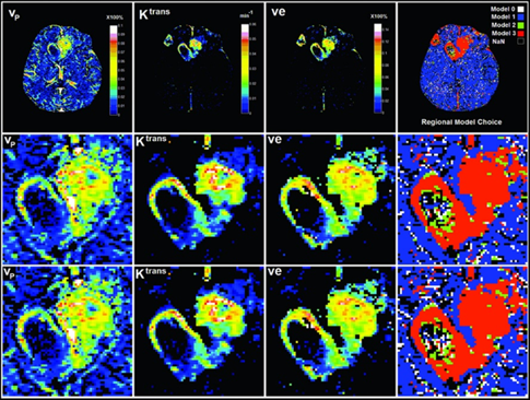
All basic MRI for anatomical images (like T2WI, T1WI, GRE, etc), Neuro perfusion, DSC-based perfusion, DCE-based perfusion and permeability, Cerebral blood flow (2D and 3D ASL), Advanced less-distorted DWI, Selective excitement. DTI (B-value ranges from 0 -10000 s/mm2, directions 256) and fMRI (NordicNeuroLab fMRI and Visual system).
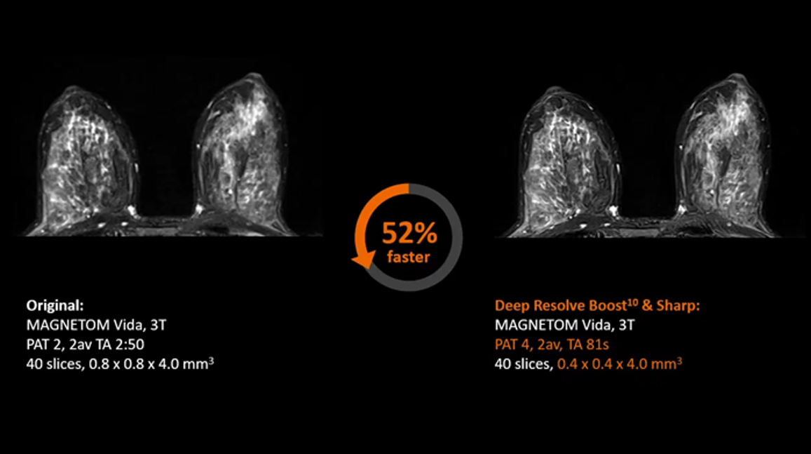
点击图片查看大图和说明

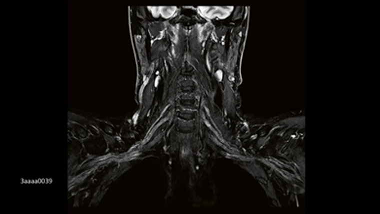
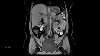
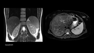
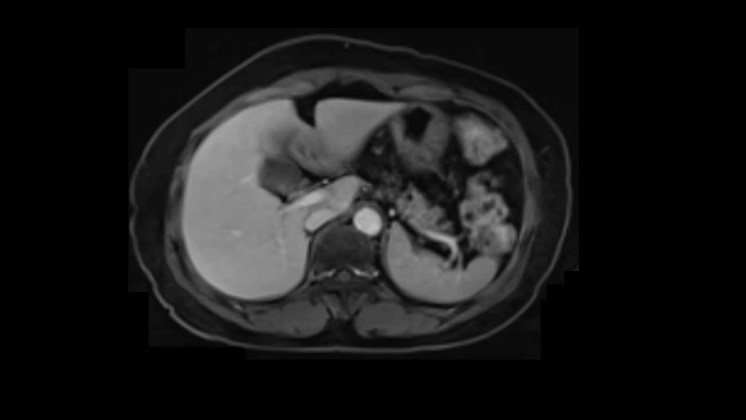
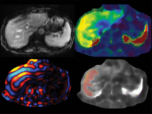
有或没有脂肪抑制的解剖成像的所有标准序列。 All advanced sequences for liver imaging for determining fat and iron, MR Elastrography to determine liver fibrosis.
点击图片查看大图和说明
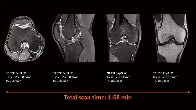
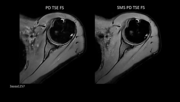
基本序列,T1, T2, T2*映射。 WARP和Advanced WARP(金属植入物)。
点击图片查看大图和说明
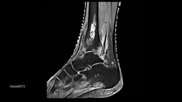
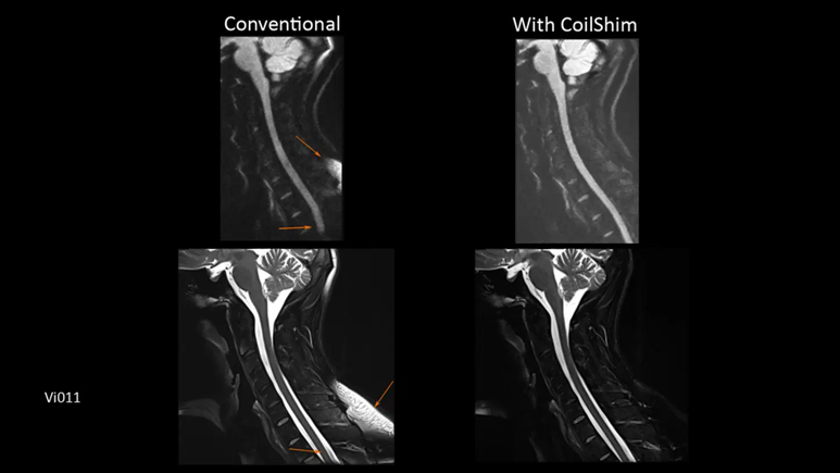
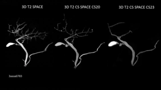
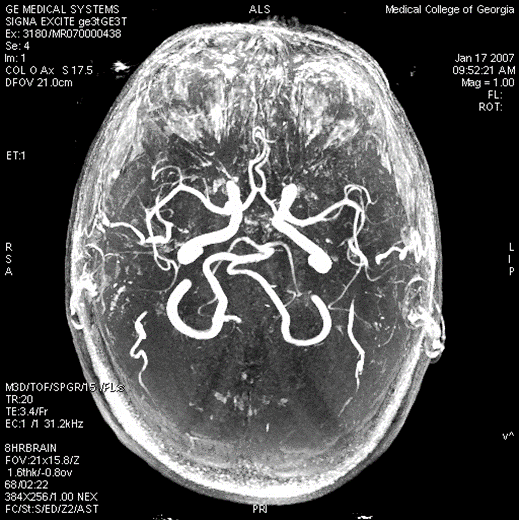
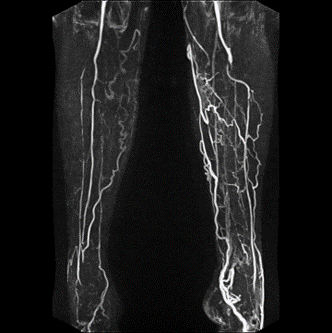
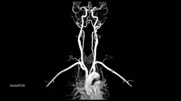
对比前后的MRA。 对比后多相MRA。 MR-DSA。 Arterial wall thickness and atherosclerotic plaques can be determined.
点击图片查看大图和说明

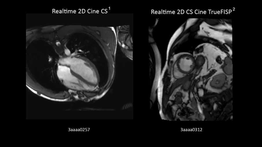
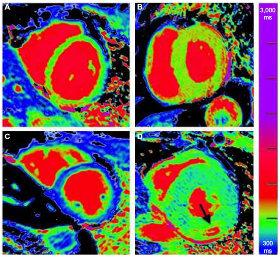
MRI is considered gold standard for myocardial thickness and changes in the relaxivity due to chemotherapy and other injury. 可以创建T1- T2-, T2*映射。
点击图片查看大图和说明
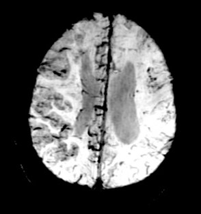
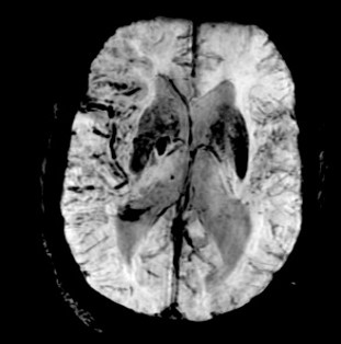
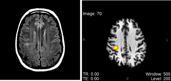
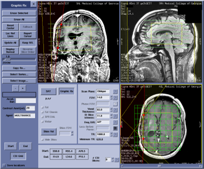
Magnetic resonance (MR) spectroscopy is a noninvasive diagnostic test for measuring biochemical changes in the brain, especially the presence of tumors. MR spectroscopy compares the chemical composition of normal brain tissue with abnormal tumor tissue.
点击图片查看大图和说明
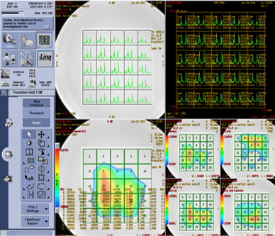
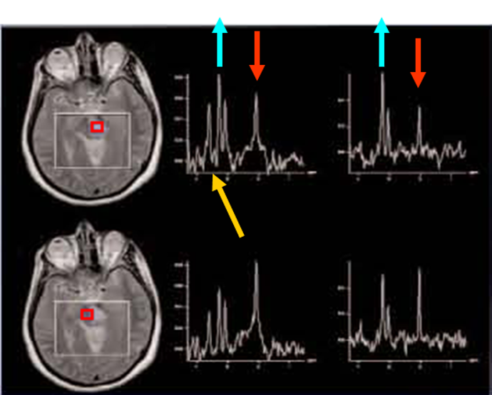
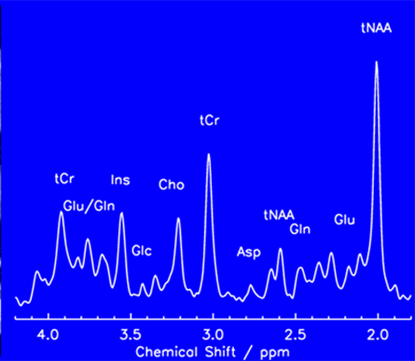
点击图片查看大图和说明